

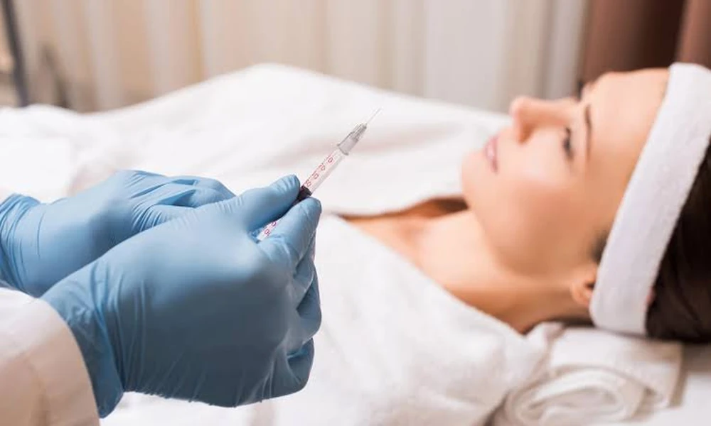
Measurement is the entire point of a before-and-after study. Skin condition is recorded at baseline, the compound is applied for a fixed period, and the same recording is repeated under identical conditions at the end. Trials involving ghk cu typically run eight to twelve weeks, since collagen turnover needs that long to produce readable movement. A shorter window captures hydration shifts at best, while structural change in the dermis stays invisible. How researchers capture that movement, and how they score it once captured, decides whether the study means anything at all, and both stages are explained directly below.
Before after imaging
Imaging in these trials begins with standardised photography. Fixed lighting, set camera distance, and controlled head position allow the same facial region to be compared across every visit, and cross-polarised filters remove surface shine so redness and pigment beneath the surface become visible. Any drift in these conditions would bury real change under ordinary shadow, which is why serious facilities photograph participants inside enclosed imaging booths rather than open rooms. Instrument scanning then goes past what photographs record. On the first and last visits, optical coherence tomography measures epidermal thickness and dermal density without cutting the skin. During the process of building new collagen, the echo strength climbs in the dermis. Profilometry converts wrinkle depth into precise numbers rather than just suggesting them with photographs alone. Corneometry probes add water content readings from the outer layer, while suction devices stretch the skin gently and time how quickly it returns, giving elasticity a numerical value. Every device produces one baseline figure and one endpoint figure, and the distance between the two is the change the study reports.
Peptide change scoring
Scoring converts raw images and readings into results that a trial can defend, and it runs on two tracks that check each other.
• Blinded human grading
Trained graders rate wrinkle depth, firmness, and tone on fixed scales without knowing which record came first or which site received treatment. Removing that knowledge removes expectation bias from the numbers. Multiple graders are usually used in protocols, since a score that only one person can reproduce is relatively ineffective. In addition to questionnaires, standardised forms allow participants to provide their subjective opinions on the smoothness and comfort of the ride, so subjective experiences are entered into the record in a controlled manner rather than as random observations.
• Statistical comparison
Treated sites are set against untreated or placebo sites on the same participant, which cancels out differences in age, diet, and sun history that comparisons between separate people would carry. A difference counts only when it clears chance at a defined threshold, so sample size and identical test conditions carry as much weight as the compound itself. Small groups measured under loose conditions generate numbers that look impressive and mean nothing, which is why published peptide trials state their participant counts and confidence levels openly. Seasonal timing gets recorded too, because skin measured in humid summer behaves differently from the same skin in dry winter.
Before and after studies measure peptide skin changes by tying controlled imaging, instrument readings, and blinded scoring to a single fixed baseline. Scanners and photographers capture the shift, graders and statistics confirm that the shift is real, and same-person comparisons prevent outside variables from making an impact. In skincare, these studies’ prominence comes from their agreement across independent methods that show the result originates from the peptide, not lighting, mood, or season.